I built this section because healthcare can feel overwhelming — especially if English isn’t your first language or no one has ever explained the basics. Here you’ll find plain-language answers to the most common questions I hear from patients every week.
-
Confusion: “If the doctor doesn’t call, I don’t have results.”
Clarity: Many offices post results to your portal first. Some only call if something is abnormal. Ask: Do you use a portal, and how do I sign up? -
Confusion: “Why do I need a referral?”
Clarity: A referral tells insurance the test is medically needed. Without it you may not be scheduled or covered. Always ask if a referral is required. -
Confusion: “No insurance means no screening.”
Clarity: Florida’s Breast and Cervical Cancer Early Detection Program offers free or low-cost screening if you qualify. Include phone numbers or links in the Resources section -
Confusion: “The imaging center will call me.”
Clarity: Sometimes the center does not call. Results go to your ordering provider and to your portal. If you do not hear back, check your portal and call your provider. -
Confusion: “New doctors will have all my old images.”
Clarity: Not always. Bring prior images or request a transfer so the radiologist can compare year to year. -
Confusion: “If I had an ultrasound, I do not need a mammogram.”
Clarity: They do different jobs. Ultrasound often looks closer after a screening mammogram. One does not replace the other. -
Confusion: “Mammograms give dangerous radiation.”
Clarity: A typical mammogram is about 0.4 mSv, roughly the natural background you get in about 7 weeks. The benefits outweigh the small risk. -
Clarity: “Benign” means not cancer. “Dense breasts” means the tissue looks white on mammograms which can hide changes. “BI-RADS” is a 0–6 number that guides next steps.
-
Clarity: Screening guidelines vary. Many public sources note regular mammograms starting at age 40 for average-risk women, with timing to be decided with your provider. The key is to screen on schedule rather than waiting for symptoms.
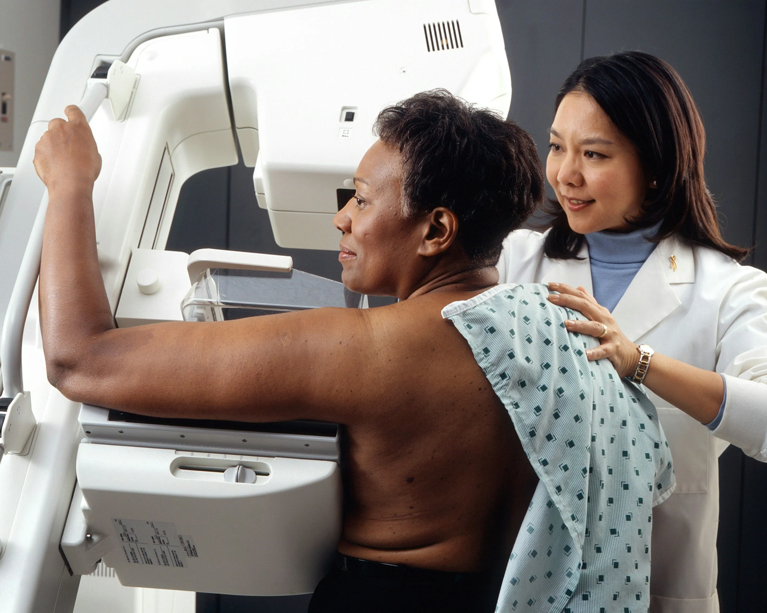
“Taking Charge of Your Breast Health”

Yearly mammograms
Radiologists compare to last year. If you skip, small changes are harder to spot.
Diagnostic mammogram
If screening shows something, this follow-up looks closer. A callback does not mean cancer.

Breast self-exam
Any age. If you feel a new lump or change, contact your provider. Waiting months can limit treatment options if something is serious..
How to read your imaging report
-
A radiology report is written by a radiologist, the doctor who interprets your imaging test. The report usually includes:
Type of exam (what test you had and when)
History/reason (why the test was ordered)
Findings (what the radiologist sees)
Impression (a summary of the key results and next steps)
The report is sent to your provider, who explains it to you. Sometimes you may see it in your health record before your provider does. If anything is unclear, ask your provider or the radiology team.
-
What BI-RADS Means
BI-RADS is a system radiologists use to describe breast imaging results (mammogram, ultrasound, MRI). It gives a number (0–6) that shows how concerning a finding is and what usually happens next.0: Incomplete — more imaging needed
1: Negative — normal, routine screening
2: Benign — harmless finding, routine screening
3: Probably benign — short-term follow up (often 6 months)
4: Suspicious — biopsy usually recommended
5: Highly suggestive of cancer — biopsy strongly recommended
6: Known cancer — used after diagnosis to guide treatment
-
What “Additional Imaging,” “Follow-Up,” or “Biopsy” Mean
Ultrasound (and other imaging) reports often include next-step recommendations. Here’s what they usually mean:Additional imaging: More pictures or a different test (like mammogram or MRI) are needed for clarity.
Follow-up imaging: Low-risk finding, but checked again later (often 6–12 months) to see if it changes.
Biopsy: A tissue sample is needed to know exactly what the cells are.
Compare with prior imaging: Old scans help show if something is stable or changing.
Surveillance/monitoring: Ongoing checks when something is probably benign but worth watching.
Imaging reports aren’t written for patients they’re written for doctors. Here’s how to make sense of yours and know what comes next.
Myth vs Fact
Fact:One screening is about 0.4 mSv, similar to 7 weeks of background. Skipping screening is riskier. (American Cancer Society).
Fact: Sometimes results are only in your portal. Always check and message your provider.
Fact: Different tools. They work together. Screening mammogram is still the standard for most.
Fact: Screening is meant to catch problems before symptoms. The exact start age and frequency should be set with your provider using trusted guidelines. (CDC overview of screening). CDC
Myth: Mammograms give too much radiation.
Myth: If no one calls, the results are fine.
Myth: Ultrasound replaces mammogram.
Myth: Screening is only for people with symptoms.